|
|
CSFoma
Peritoneal Cerebrospinal Fluid Pseudocyst
General Considerations
- Loculation of cerebrospinal fluid (CSF) at the distal end of the a ventriculoperitoneal shunt tube
- May occur secondary to adhesions
- Leads to an enlarging abdominal and/or pelvic mass at catheter tip
- CSF from tip is no longer absorbed by peritoneum
Clinical Findings
- Local signs of an abdominal mass
- Increased intracranial pressure signs
Imaging Findings
- Diagnosis is most often made by ultrasound
Treatment
- Can resolve spontaneously
- Shunt revision and repositioning
- Aspiration of cyst’s contents
Complications
- Infection of pseudocyst can reach >76% in children under 4 years
- Overall infection rate is 30%


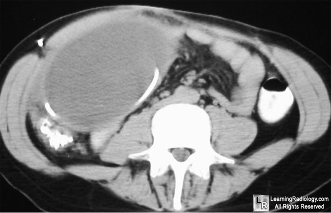
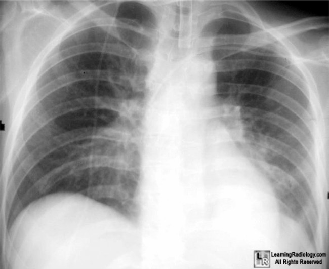
CSFoma. CT scan of lower abdomen at left shows a large fluid-filled cystic mass (black arrow) at the termination of the coiled ventriculoperitoneal shunt tube (red arrows) seen traversing the chest in the chest radiograph at right (white arrows).
For more information, click on the link if you see this icon 
For this same photo without the annotations, click here or here
Radiology of the Postoperative GI Tract
By Bruce R. Javors, Ellen L. Wolf
Edition: illustrated
Published by Springer, 2003
|
|
|
